A Different View of Your Breast Health
Non-invasive. No radiation. No compression.
There’s More to Breast Health Than Looking for a Tumor.
Most breast cancer screening focuses on structure—looking for physical abnormalities that can be seen on an image. That information is incredibly valuable, but it’s only one way of looking at breast health.
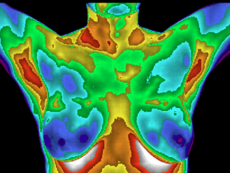
Thermography looks at something different: function. It measures temperature patterns at the skin's surface and allows those patterns to be tracked for stability or change over time.
The two technologies answer different questions—and understanding that difference is key to understanding the value of thermography.
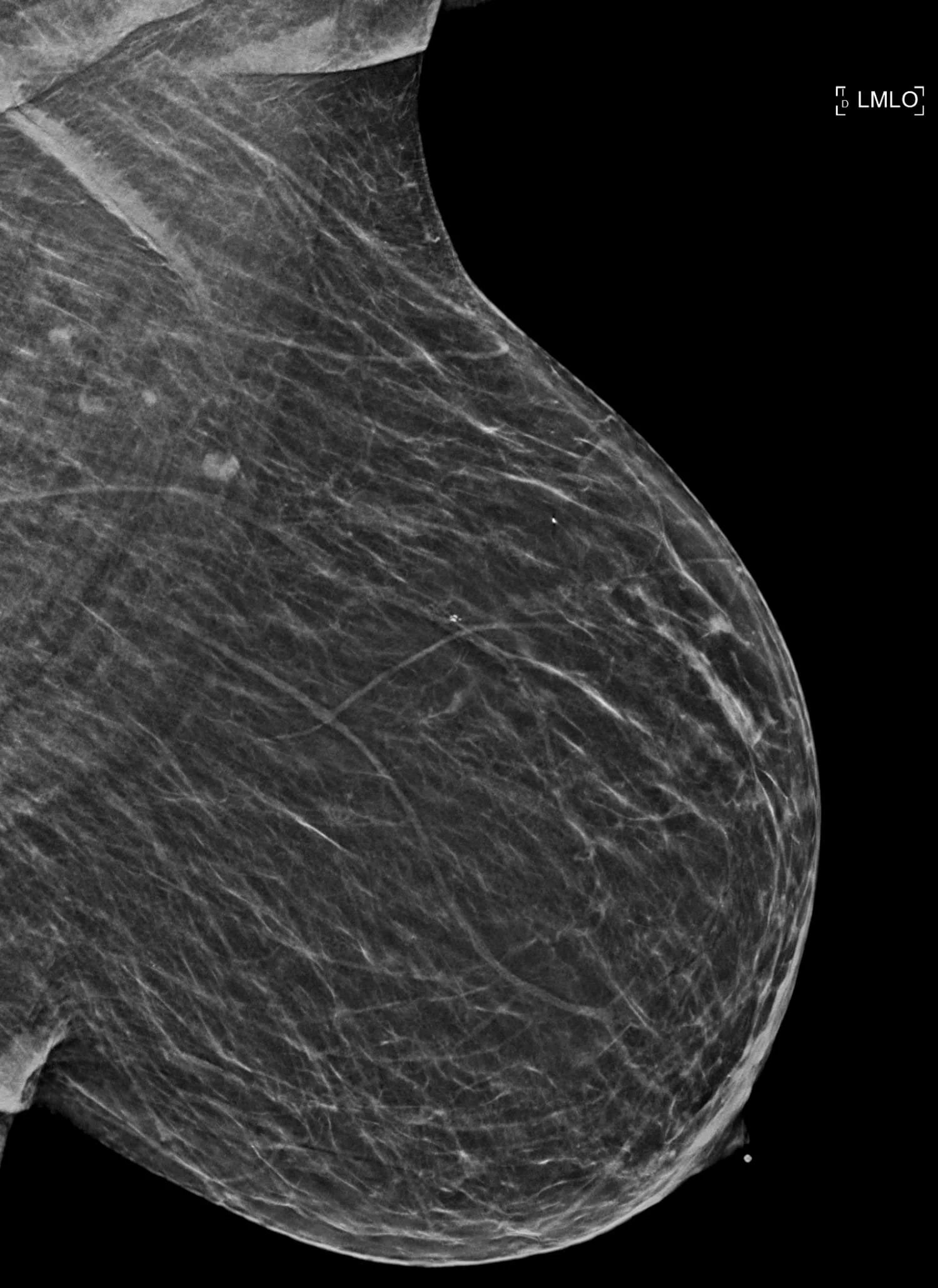
Mammography
Looks at Structure
Mammography uses low-dose X-rays to create images of breast tissue, looking for structural findings such as masses, calcifications, and other abnormalities that may require further evaluation.
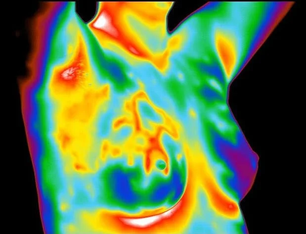
Thermography
Looks at Function
Thermography uses infrared imaging to measure and map temperature patterns at the surface of the breasts. Serial studies allow those patterns to be compared over time for stability or meaningful change.
One doesn't replace the other. They provide different information about breast health.
If structural imaging is the only tool you're using, you may be seeing only part of the picture.
Why Function?
Disease doesn't just change what tissue looks like. It can change how tissue behaves.
Abnormal tissue can have altered metabolism. Cancer cells can undergo significant metabolic changes as tumors develop and progress.
Growing tumors can develop abnormal blood supply. Angiogenesis—the formation of new blood vessels—is a well-established hallmark of cancer and an important feature of breast-cancer progression.
Metabolism and blood flow influence heat. Those physiologic processes can contribute to differences in heat reaching the skin's surface—the very patterns thermography is designed to measure.
Thermography gives us a way to follow those patterns. When thermal findings are asymmetric, unusually prominent, or change over time, they provide physiologic information that can be considered alongside structural imaging and other clinical information.
So, What Can Thermography Add?
-
A single thermogram provides a snapshot. Serial studies allow your thermal patterns to be compared over time, helping identify whether your individual pattern remains stable or meaningfully changes.
Instead of simply waiting for something to be found, you can actively follow your breast health over time.
-
Thermography is completely non-contact. Images are captured without compressing the breasts or exposing them to ionizing radiation, making repeat imaging simple and comfortable.
-
Because thermography measures surface temperature patterns rather than creating an X-ray image of breast tissue, breast density does not obscure the thermal image in the same way it can affect mammographic interpretation.
Thermography can also be performed in women with breast implants and following many breast procedures, although individual circumstances may affect interpretation.
Women deserve more than a wait-and-see approach to breast health.
From Passive Screening to Proactive Breast Health
Traditional breast screening is designed primarily to look for structural signs of disease. That’s important—but being proactive about breast health can mean looking beyond a single point-in-time image.
With thermography, we begin by establishing your individual thermal baseline. Future studies can then be compared with that baseline to see whether your thermal patterns remain stable or change over time.
The goal isn’t simply to take a thermogram. It’s to understand your pattern, follow it over time, and know when a change deserves attention.
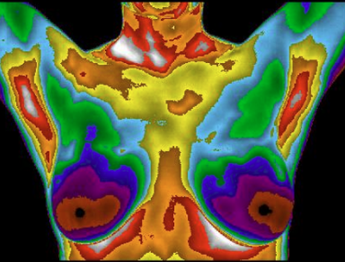
01 Document Your Pattern
Your initial thermogram documents your individual thermal pattern.
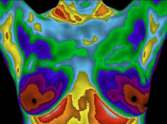
02 Establish Your Baseline
A follow-up study confirms whether your thermal pattern remains stable.
03 Track Over Time
Periodic studies allow your established baseline to be monitored for meaningful change.
Your breast health isn't a single image. It's a story that unfolds over time.
What Our Clients Are Saying

“Had initial Thermograph in January 2025. Everyone at the clinic was so nice and helpful. Dr. Baggett explained everything thoroughly and then followed up with results in detail. I would highly recommend them for your Thermology Imaging needs!”
— Alesia, Google Review“Mrs. Jo is the Best at what she does; she is caring and makes sure you feel comfortable. Dr Blake is professional and makes sure to answer any questions you might have.”
— Vickie, Google Review“I love the holistic approach Dr. Blake’s thermography service provides to women’s healthcare. I had my first appointment yesterday, and Jo is definitely a people person who cares about the patients. I eagerly await my results and recommend this office to anyone and everyone! They make you feel like a family!”
— Megan , Google Review
The Process Is Simple
01 - Schedule Your Visit
Choose a convenient appointment time online. If this is your first thermogram with us, select the New Client Baseline Study.

02 - Get Your Thermogram
Your imaging appointment takes about 45 minutes. There's no radiation, no compression, and no physical contact during imaging.

03 - Understand Your Results
Your images are professionally interpreted, and you'll receive your complete report along with a personal video review explaining your findings and recommended next steps.
You'll receive more than an image—you'll have a starting point for understanding and tracking your breast health over time.
Ready to Get Started?
Need help with the cost? Qualified women may be eligible for up to $150-$200 in reimbursement through the United Breast Cancer Foundation. Flexible payment options are also available.

Not Ready to Schedule Yet?
Start with something free.
Get our 6 Breast Health Tips guide and discover practical ways to take a more proactive approach to your breast health.